Fundamentals of EEG in Epilepsy and Neurological Assessment
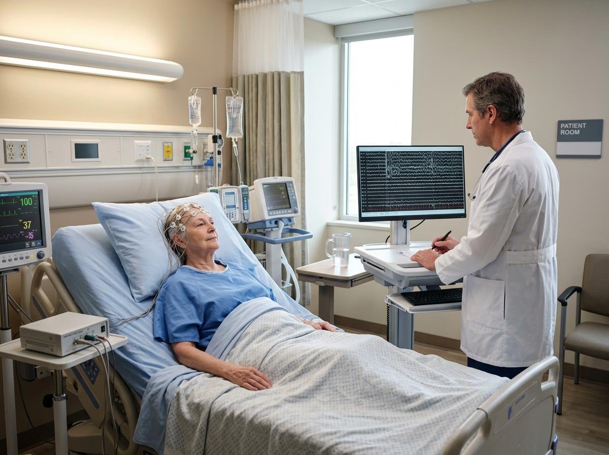
Epilepsy diagnosis can be complex. It often relies on capturing fleeting electrical signals in the brain. For nearly a century, the electroencephalogram (EEG) has been our primary window into this activity. Discovered by Hans Berger in 1929, the EEG revolutionized our understanding of cortical excitability.
Yet, traditional EEG methods sometimes face challenges. Access to specialized neurodiagnostic care can be limited, especially in underserved regions. This creates a diagnostic gap for many patients.
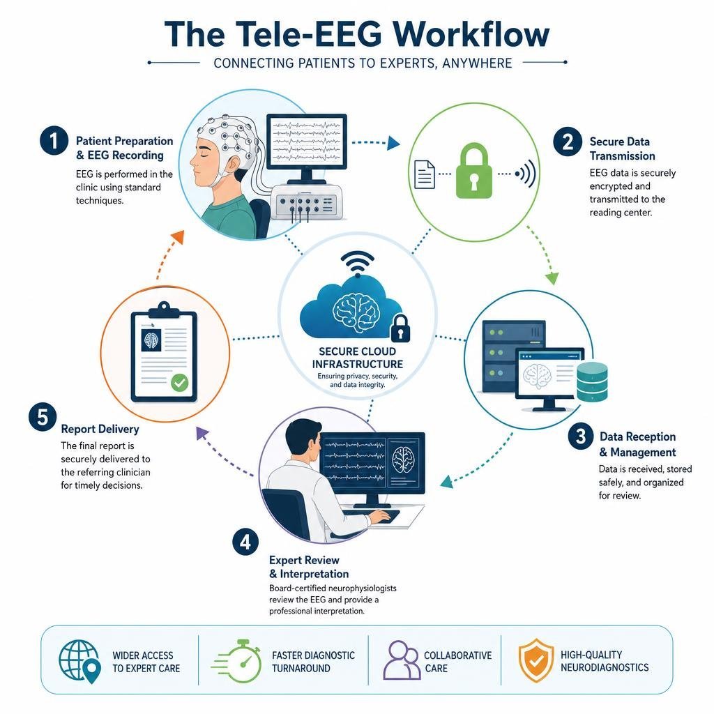
This is where Tele-EEG steps in. By leveraging digital technology, we can now provide remote interpretation and faster diagnostic turnaround. This ensures more patients receive timely and accurate epilepsy diagnoses. For instance, advanced platforms facilitate remote continuous EEG monitoring, allowing specialists to review data from anywhere.
In this comprehensive guide, we will explore the fundamentals of EEG. We will cover how it works, its different types, and its crucial role in diagnosing epilepsy and other neurological conditions. We will also delve into the transformative power of Tele-EEG. This includes its clinical applications, diagnostic waveform patterns, and answers to frequently asked questions.
The electroencephalogram (EEG) functions as a non-invasive tool to record the electrical activity generated by the brain. This activity primarily originates from the synchronized postsynaptic potentials of pyramidal neurons located in the cerebral cortex. When thousands of these neurons fire in concert, their electrical signals are strong enough to be detected by electrodes placed on the scalp.
To ensure consistency and comparability across recordings, a standardized electrode placement system, known as the 10-20 system, is universally employed. This system dictates the precise locations for electrodes on the scalp, based on anatomical landmarks, ensuring that specific brain regions are consistently monitored. The electrodes, typically small metal discs, are affixed to the scalp with a conductive paste, picking up the minute voltage fluctuations that reflect brain activity. These signals are then amplified and displayed as waveforms or “traces” on a monitor, allowing neurologists to interpret the brain’s ongoing electrical rhythms.
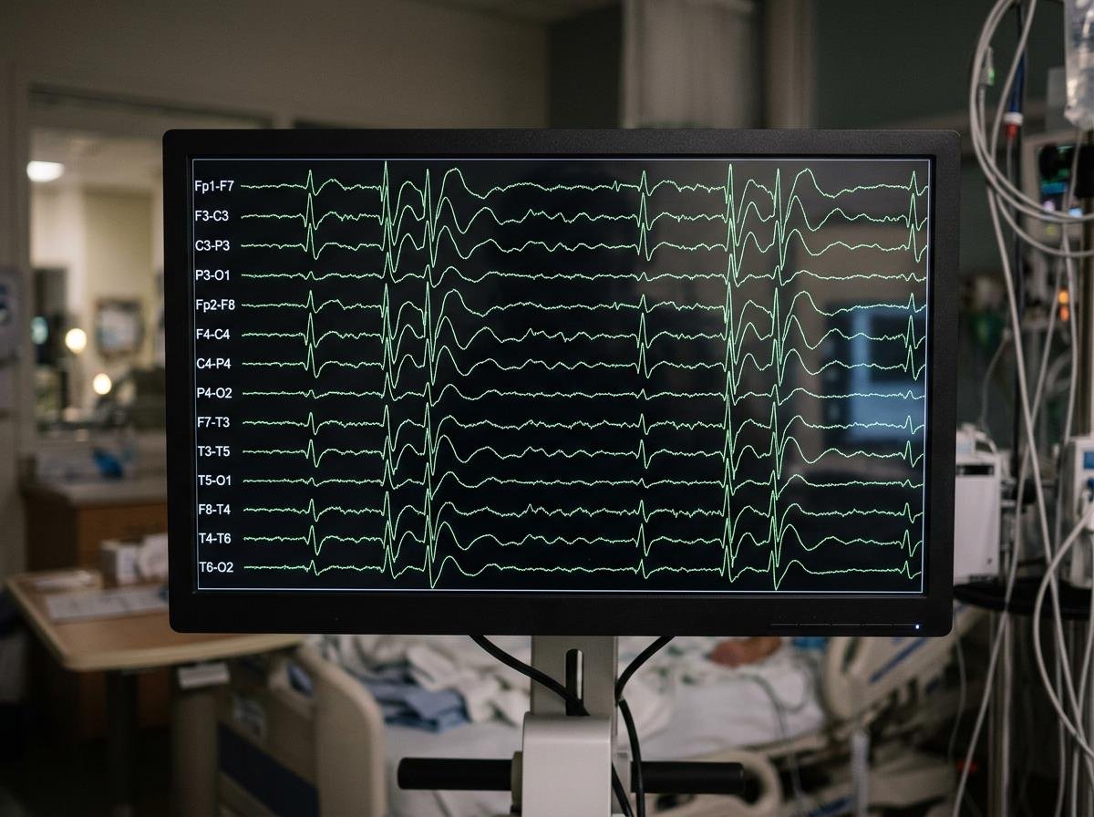
In the context of epilepsy, the primary diagnostic markers on an EEG are interictal epileptiform discharges (IEDs). These are transient, abnormal electrical events that occur between seizures. IEDs represent sudden, excessive, and hypersynchronous neuronal activity. They manifest as distinctive patterns on the EEG, most commonly as “spikes” or “sharp waves.” Spikes are characterized by a very rapid rise and fall, typically lasting less than 70 milliseconds, while sharp waves have a slightly broader duration, ranging from 70 to 200 milliseconds, with a somewhat blunter appearance. The presence, morphology, location, and frequency of these IEDs are critical indicators of an underlying epileptogenic process.
To standardize the identification and interpretation of these crucial findings, the International Federation of Clinical Neurophysiology (IFCN) has established specific criteria. These guidelines help clinicians differentiate true epileptiform activity from benign variants or artifacts, ensuring more accurate diagnoses. However, IEDs are not always present in individuals with epilepsy, nor do they exclusively occur in those with the condition; between 1 and 13 percent of normal individuals can show IEDs on EEG. Despite this, IEDs remain the most indicative diagnostic finding for epilepsy, providing objective evidence of cortical hyperexcitability.
Activation Procedures and Diagnostic Sensitivity
A routine EEG recording, typically lasting 20 to 40 minutes, may not always capture epileptiform activity, especially if seizures are infrequent or the IEDs are sporadic. To increase the diagnostic yield and provoke latent abnormalities, neurologists often employ various activation procedures during an EEG. These techniques are designed to stress the brain or alter its physiological state, making epileptiform activity more likely to emerge.
One common activation procedure is hyperventilation. Patients are instructed to breathe deeply and rapidly for several minutes. This maneuver leads to a decrease in carbon dioxide levels in the blood, causing cerebral vasoconstriction and a temporary reduction in blood flow to the brain. This physiological change can often trigger or exacerbate certain types of epileptiform discharges, particularly generalized spike-and-wave discharges characteristic of absence seizures. For example, a child with suspected absence seizures might show classic 3-Hz generalized spike-wave discharges only during hyperventilation. Clinicians may even ask patients to count their breaths to monitor for subtle cognitive impairment during these events.
Intermittent photic stimulation (IPS) involves exposing the patient to a series of flashing lights at varying frequencies. This procedure is particularly useful for identifying photosensitive epilepsy, a condition where seizures are triggered by specific visual stimuli. About 3% of people with epilepsy are affected by photosensitivity, and IPS can reliably provoke photoparoxysmal responses on the EEG in these individuals.
Sleep deprivation is another powerful activation method. Patients are asked to reduce their sleep significantly the night before the EEG. Drowsiness and sleep, especially during the transition phases, are well-known to activate epileptiform discharges that might be absent during wakefulness. This is because sleep alters brain rhythms and neuronal excitability. Partial sleep deprivation, where patients get only a few hours of sleep, has been shown to be as effective as complete all-night deprivation and is often more patient-friendly. This technique is especially valuable for capturing nocturnal seizures or IEDs that are sleep-dependent.
These activation procedures significantly enhance the diagnostic sensitivity of EEG, helping to reveal subtle baseline abnormalities or provoke reflex seizures that might otherwise be missed. By strategically utilizing these techniques, clinicians can substantially increase the chances of capturing crucial electrographic evidence necessary for an accurate epilepsy diagnosis.
Differentiating Epileptic Seizures from Nonepileptic Events
One of the most challenging aspects of epilepsy diagnosis is distinguishing true epileptic seizures from other paroxysmal events that mimic them. These “nonepileptic events” can include a wide range of conditions, from cardiovascular issues like syncope to psychological phenomena such as psychogenic nonepileptic spells (PNES). Accurate differentiation is paramount, as misdiagnosis can lead to inappropriate and potentially harmful treatments, delayed effective care, and significant psychological distress for the patient.
Psychogenic nonepileptic spells (PNES) are behavioral events that resemble epileptic seizures but are not caused by abnormal electrical activity in the brain. Instead, they are typically a manifestation of psychological distress. PNES account for a substantial proportion of patients seen in epilepsy centers, with approximately 30% of admissions to epilepsy monitoring units attributed to these spells. Differentiating PNES from epileptic seizures relies heavily on correlating clinical semiology (the observable signs and symptoms of the event) with simultaneous EEG recordings. During an epileptic seizure, the EEG will show characteristic ictal (seizure-related) patterns, whereas during a PNES event, the EEG typically remains normal or shows non-specific changes unrelated to epileptiform activity.
Other conditions that can be mistaken for epilepsy include syncope (fainting due to temporary reduction in blood flow to the brain), transient ischemic attacks (TIAs), migraines with aura, movement disorders, and sleep disorders. While clinical history and witness accounts are crucial, definitive differentiation often requires objective electrophysiological evidence.
This is where video-EEG monitoring becomes indispensable. By combining continuous EEG recording with synchronized video capture, clinicians can precisely correlate a patient’s observed behavior during an event with the underlying brain electrical activity. This powerful tool allows neurologists to see exactly what the patient is doing during a spell and simultaneously analyze their brainwaves. For instance, a 32-year-old man presenting with sudden falls might be suspected of having focal seizures, but video-EEG could reveal that these are actually syncopal episodes with secondary anoxic seizures, or even PNES.
The ability of video-EEG to provide this comprehensive, real-time correlation is why it leads to altered diagnoses in 45% to 58% of patients and changes in management in nearly 75% of monitored patients. This high diagnostic yield underscores its critical role in avoiding misdiagnosis, ensuring that patients receive the correct diagnosis and, consequently, the most appropriate and effective treatment for their condition.
Expanding Clinical Reach: Tele-EEG and Neurodiagnostics in Practice
The advent of digital technology has profoundly transformed neurodiagnostics, particularly with the rise of Tele-EEG. Tele-EEG refers to the remote acquisition, transmission, and interpretation of EEG data, effectively extending the reach of specialized neurological expertise beyond traditional hospital settings. This innovation addresses geographical barriers and staffing shortages, bringing high-quality neurodiagnostic services to a wider patient population.
One of the most significant advancements in this area is remote continuous EEG monitoring. This technology allows neurologists and neurophysiologists to review EEG data in real-time or near real-time from virtually any location, whether it’s a critical care unit, a patient’s home, or an outpatient clinic. Platforms like those offered by CortiCare facilitate this seamless remote access, enabling expert interpretation and timely decision-making. This capability is especially vital in situations requiring immediate intervention, such as in the intensive care unit (ICU).
In the ICU, continuous EEG monitoring is a game-changer. Critically ill patients, particularly those with altered mental status, are at high risk for non-convulsive seizures or non-convulsive status epilepticus, conditions that are often clinically subtle or entirely invisible without continuous brain activity monitoring. Tele-EEG allows for constant surveillance of these patients, enabling prompt detection and treatment of seizures, which can significantly impact neurological outcomes. For example, monitoring anesthesia depth during medically induced coma also benefits from continuous EEG, ensuring optimal patient management.
The decentralized access offered by Tele-EEG is revolutionizing neurodiagnostic care. It means that rural hospitals or smaller clinics, which may lack on-site neurophysiology specialists, can still provide advanced EEG services. Data can be transmitted securely to expert centers, where experienced neurologists can perform real-time review and provide immediate feedback or interpretations. This not only improves patient safety and care quality but also reduces the need for patient transfers, saving time and resources. The ability to conduct real-time review ensures that critical changes in brain activity are identified swiftly, leading to faster diagnoses and more effective management strategies.
Clinical Applications of Tele-EEG and Neurodiagnostics in Ambulatory Settings
Beyond the critical care environment, Tele-EEG has expanded the utility of EEG into ambulatory settings, offering a flexible and patient-centric approach to long-term monitoring. Ambulatory EEG involves patients wearing a portable EEG device, often for extended periods ranging from 24 to 72 hours, or even longer. This allows for continuous recording of brain activity while the patient goes about their normal daily routines, often in their home environment.
The primary advantage of ambulatory EEG is its ability to capture infrequent or subtle events that might be missed during a short, routine EEG performed in a clinic. Since many seizures are unpredictable and sporadic, monitoring over several days significantly increases the chance of capturing a habitual spell. While traditional ambulatory EEG may not always include synchronized video, patients are typically asked to keep a detailed diary of their activities and any experienced symptoms, aiding in behavioral correlation. Some advanced ambulatory systems now offer integrated video, further enhancing their diagnostic power by linking observed behavior directly to EEG changes.
The sheer volume of data generated by 24-72 hour tracings necessitates sophisticated analysis tools. Automated spike detection software helps clinicians sift through vast amounts of EEG data, flagging potential interictal epileptiform discharges or seizure patterns for expert review. While these algorithms are powerful, human oversight remains crucial to differentiate true epileptiform activity from artifacts and benign variants, ensuring diagnostic accuracy.
Ambulatory Tele-EEG is particularly beneficial for patients whose seizures are infrequent, nocturnal, or occur only in specific real-world contexts. It offers increased patient comfort and convenience compared to inpatient video-EEG monitoring, as patients can remain in their familiar surroundings. However, it’s important to understand the distinctions between different EEG modalities:
| Modality | Duration | Primary Indication | Key Feature |
|---|---|---|---|
| Routine EEG | 20-40 minutes | Initial assessment for suspected epilepsy, general brain activity evaluation. | Quick, non-invasive, captures immediate brain state. |
| Ambulatory EEG | 24-72+ hours | Capturing infrequent seizures, monitoring nocturnal events, assessing seizure frequency in natural environment. | Portable, records outside clinic, increased chance of capturing sporadic events. |
| Inpatient Video-EEG | Days to weeks | Definitive seizure characterization, differentiating epileptic from non-epileptic events, presurgical evaluation. | Continuous EEG with synchronized video, direct medical supervision, allows for seizure provocation (e.g., AED withdrawal). |
Presurgical Evaluation and Advanced Tele-EEG and Neurodiagnostics
For individuals with drug-resistant epilepsy—meaning their seizures are not controlled by anti-seizure medications—epilepsy surgery can offer a chance at seizure freedom. A critical step in this process is the presurgical evaluation, which aims to precisely localize the seizure onset zone (SOZ), the small area of the brain where seizures originate. While non-invasive EEG and imaging (like MRI and PET) provide valuable clues, sometimes more advanced, invasive techniques are required to pinpoint the SOZ with sufficient accuracy for surgical resection.
In these complex cases, invasive EEG monitoring, often referred to as Phase II monitoring, is employed. This involves surgically placing electrodes directly on or within the brain. Two primary types of invasive EEG are utilized:
- Stereo-EEG (sEEG): This technique involves implanting thin, multi-contact electrodes deep into the brain along a stereotactic trajectory. sEEG provides a three-dimensional mapping of brain activity, allowing for precise localization of deep-seated or widely distributed seizure foci. It is particularly useful when non-invasive studies yield ambiguous or bilateral findings. However, research indicates that for patients with bilateral or unclear scalp EEG onset, the diagnostic yield of sEEG for identifying a single, resectable SOZ can be limited, with only about 14% of such cases identifying a single SOZ. This highlights the importance of careful patient selection and integration with other multimodal data.
- Subdural grids and strips: These are flexible sheets or strips of electrodes that are placed directly on the surface of the brain (the cerebral cortex) after a craniotomy. They provide excellent spatial resolution for cortical areas and are often used when the SOZ is suspected to be superficial.
During invasive EEG monitoring, patients remain in a specialized epilepsy monitoring unit, where their brain activity is continuously recorded, often for days or weeks. This allows clinicians to capture multiple habitual seizures and precisely map their origin and spread. The data from invasive EEG is frequently integrated with multimodal MRI fusion, combining anatomical imaging with functional data to create a comprehensive picture of the brain’s structure and function in relation to the seizure focus.
The ultimate goal of this rigorous evaluation is to perform surgical triage—determining if a patient is a suitable candidate for resective surgery and, if so, guiding the neurosurgeon to the exact area to be removed. Post-surgical outcomes are typically assessed using the Engel classification, a widely accepted scale that categorizes seizure freedom and reduction. While invasive EEG is a powerful tool, it’s a significant undertaking, and patients are carefully selected based on the likelihood of achieving a favorable outcome.
Diagnostic Waveform Patterns Across Specific Epilepsy Syndromes
The EEG is not just a tool for detecting seizures; it’s also crucial for classifying the specific type of epilepsy and identifying distinct epilepsy syndromes. Many epilepsy syndromes are characterized by unique and recognizable EEG patterns, which are vital for guiding prognosis and selecting the most effective anti-seizure medications. Recognizing these characteristic waveforms is a cornerstone of neurodiagnostic interpretation.
One of the most classic examples is Childhood Absence Epilepsy (CAE). This syndrome is defined by brief, sudden lapses of consciousness, often accompanied by a distinctive EEG signature: generalized, symmetrical, and synchronous 3-Hz spike-and-wave discharges. These discharges typically begin and end abruptly, correlating directly with the clinical absence seizure. The regularity and frequency of these discharges are highly characteristic and help differentiate CAE from other types of absence seizures or generalized epilepsies.
Juvenile Myoclonic Epilepsy (JME), another common generalized epilepsy syndrome, also presents with a characteristic EEG pattern. Patients with JME typically exhibit generalized polyspike-and-wave discharges, which are often faster than the 3-Hz seen in CAE, ranging from 3.5 to 6 Hz. These discharges are often provoked by sleep deprivation or photic stimulation and are highly indicative of JME, informing treatment strategies that prioritize drugs effective against generalized epilepsies.
In infancy, West Syndrome, characterized by infantile spasms, developmental regression, and a highly chaotic EEG pattern called hypsarrhythmia, is a severe epilepsy syndrome. Hypsarrhythmia is a high-amplitude, disorganized, and multifocal pattern with continuous slow waves and multifocal spikes and sharp waves. This pattern is pathognomonic for West Syndrome and demands urgent diagnosis and specific treatment approaches.
Another severe childhood epilepsy, Lennox-Gastaut Syndrome (LGS), is associated with multiple seizure types and cognitive impairment. Its characteristic EEG finding is a generalized slow spike-and-wave pattern, typically occurring at a frequency of 1.5 to 2.5 Hz. This slower frequency differentiates it from the faster spike-and-wave patterns seen in other generalized epilepsies and is a key diagnostic criterion for LGS.
Recognizing these diagnostic syndromic EEG traces is not merely an academic exercise; it has profound implications for patient care. An accurate syndromic diagnosis allows clinicians to:
- Provide a precise prognosis: Different syndromes have varying long-term outcomes.
- Select appropriate medications: Certain anti-seizure drugs are highly effective for specific syndromes, while others can worsen seizures.
- Guide management strategies: This includes counseling, lifestyle adjustments, and potential surgical considerations.
- Avoid misdiagnosis: Preventing the use of ineffective or harmful treatments.
The EEG, therefore, serves as an invaluable roadmap for navigating the complex landscape of epilepsy, enabling tailored and effective interventions.
Frequently Asked Questions About Tele-EEG and Epilepsy Diagnosis
Can a normal routine EEG rule out an epilepsy diagnosis?
No, a normal routine EEG, while a valuable diagnostic tool, absolutely cannot definitively rule out an epilepsy diagnosis. This is a critical point that patients and clinicians must understand. Several factors contribute to this limitation:
Firstly, intermittent discharges are a common characteristic of epilepsy. Epileptiform activity, such as spikes and sharp waves, often occurs sporadically between seizures (interictally) rather than continuously. A brief, routine EEG recording, typically lasting only 20 to 40 minutes, has a limited chance of capturing these transient events. It’s like trying to catch a fleeting moment with a very short camera exposure.
Secondly, temporal sampling is a major constraint. With such a short recording window, the probability of capturing an actual seizure (ictal event) or even significant interictal activity is inherently low, especially if seizures are infrequent. Even in patients with confirmed epilepsy, the first routine EEG may be normal in a substantial minority.
Thirdly, spatial sampling can be an issue. Routine scalp EEG electrodes primarily detect activity from the superficial cortical areas. Epileptiform discharges originating from deeper brain structures, such as the mesial temporal lobe, or from basal cortical regions, may not be adequately picked up by scalp electrodes. This can lead to false-negative results.
Therefore, if clinical suspicion for epilepsy remains high despite a normal routine EEG, further investigations are often warranted. These may include repeated recordings, often after sleep deprivation, or more prolonged monitoring such as ambulatory telemetry or inpatient video-EEG. These longer recordings significantly increase the temporal sampling, improving the likelihood of capturing epileptiform activity or a clinical event. The clinical history and semiology remain paramount, and EEG findings must always be interpreted within that broader clinical context.
How does tele-EEG compare to standard in-hospital video-EEG monitoring?
Tele-EEG and standard in-hospital video-EEG monitoring both aim to provide comprehensive insights into brain electrical activity for epilepsy diagnosis, but they differ significantly in their operational models and specific advantages.
Standard in-hospital video-EEG monitoring is considered the gold standard for definitive seizure characterization and presurgical evaluation. Patients are admitted to an epilepsy monitoring unit (EMU) for days to weeks, where they are continuously monitored by dedicated staff. The key advantage is the synchronized video and EEG, allowing precise correlation of clinical behavior with brain electrical activity, and the ability for immediate medical intervention during seizures or for controlled anti-seizure medication withdrawal.
Tele-EEG, on the other hand, leverages technology to facilitate remote access and interpretation. While some Tele-EEG setups can mirror inpatient video-EEG in critical care settings, the term often refers to remote review of data acquired either in an outpatient clinic or through ambulatory monitoring at home.
Here’s a comparison:
- Remote Transmission & Cloud Architecture: Tele-EEG excels here. Data is transmitted digitally, often to a secure cloud platform, allowing neurophysiologists to review studies from anywhere. This overcomes geographical barriers, bringing expert interpretation to facilities without on-site specialists. In-hospital monitoring typically involves local data acquisition and review within the facility.
- Patient Comfort & Convenience: Ambulatory Tele-EEG significantly enhances patient comfort by allowing monitoring in the patient’s natural home environment, reducing the need for lengthy hospital stays. In-hospital monitoring, while medically comprehensive, requires patients to be confined to a hospital room.
- Cost Efficiency: Tele-EEG, particularly ambulatory forms, can be more cost-efficient by reducing inpatient days, associated hospital costs, and travel expenses for patients and their families. This makes advanced neurodiagnostic services more accessible.
- Rapid Interpretation: Tele-EEG can often provide faster access to expert interpretation. Once data is transmitted, a remote specialist can review it promptly, potentially shortening diagnostic delays. In-hospital monitoring interpretation is also rapid for critical events but overall reporting may follow standard hospital workflows.
- Clinical Supervision & Intervention: In-hospital video-EEG offers immediate medical supervision and intervention during seizures, which is crucial for patient safety, especially during medication withdrawal. While Tele-EEG in critical care can provide real-time alerts, ambulatory Tele-EEG typically lacks immediate medical oversight for severe events.
- Behavioral Correlation: In-hospital video-EEG provides superior, high-fidelity synchronized video for precise behavioral-EEG correlation. Ambulatory Tele-EEG may rely on patient diaries or less sophisticated integrated video, potentially limiting the detail of behavioral correlation.
Tele-EEG expands the reach and accessibility of neurodiagnostic services, offering flexibility and efficiency, particularly in ambulatory and critical care settings. Standard in-hospital video-EEG remains the gold standard for complex cases requiring intensive, supervised monitoring and precise behavioral-EEG correlation for presurgical evaluation or challenging differential diagnoses. The choice between them depends on the specific clinical question, patient needs, and available resources.
Why are interictal epileptiform discharges crucial for treatment planning?
Interictal epileptiform discharges (IEDs), the abnormal electrical spikes and sharp waves observed on an EEG between seizures, are far more than just a diagnostic curiosity; they are crucial biomarkers that profoundly influence treatment planning for individuals with epilepsy.
Firstly, IEDs serve as a direct epileptogenicity biomarker. Their presence on an EEG provides objective evidence of abnormal cortical excitability, indicating that the brain has a predisposition to generate seizures. This electrophysiological signature confirms the underlying pathological process of epilepsy, even if a seizure has not been captured during the recording.
Secondly, IEDs are powerful predictors of seizure recurrence risk. For patients who have experienced a first unprovoked seizure, the presence of IEDs significantly increases the likelihood of future seizures. Similarly, in individuals with established epilepsy, the frequency and morphology of IEDs can inform decisions about anti-seizure medication (ASM) withdrawal; a high burden of IEDs often suggests a higher risk of seizure relapse if medications are tapered.
Thirdly, the characteristics of IEDs directly guide medication selection. Different types of IEDs (e.g., focal vs. generalized, specific patterns like 3-Hz spike-wave) correlate with distinct epilepsy types and syndromes. For instance, generalized spike-and-wave discharges suggest a generalized epilepsy, which responds best to certain ASMs (e.g., valproate, lamotrigine) but can be worsened by others (e.g., carbamazepine). Conversely, focal IEDs point towards focal epilepsy, for which other ASMs are more appropriate. Accurate identification of these patterns helps neurologists tailor medication regimens, maximizing efficacy and minimizing adverse effects.
Fourthly, IEDs are fundamental for syndrome identification. As discussed, specific IED patterns (e.g., hypsarrhythmia, slow spike-wave) are pathognomonic for severe epilepsy syndromes like West Syndrome or Lennox-Gastaut Syndrome. Identifying these syndromes through EEG is critical because they dictate not only specific treatment protocols but also provide important prognostic information regarding cognitive and developmental outcomes.
Finally, monitoring IEDs can be valuable for baseline tracking and assessing treatment effectiveness. While not a perfect correlation, a significant reduction or disappearance of IEDs on follow-up EEGs can sometimes indicate a positive response to ASM therapy, offering supportive evidence for treatment adjustments.
In summary, IEDs provide invaluable electrophysiological insights that move beyond mere diagnosis. They inform risk stratification, guide pharmacotherapy, enable precise syndromic classification, and contribute to the ongoing management of epilepsy, making them an indispensable component of comprehensive treatment planning.
Conclusion
The electroencephalogram (EEG) has stood the test of time as an indispensable cornerstone in the diagnosis and management of epilepsy. From Hans Berger’s initial discovery to today’s sophisticated digital platforms, EEG continues to offer a unique window into the brain’s electrical landscape. We’ve explored its fundamental principles, the critical role of activation procedures, and its unmatched ability to differentiate epileptic seizures from their mimics.
The evolution of remote telemetry through Tele-EEG has dramatically expanded neurodiagnostic access, overcoming geographical barriers and bringing specialized care to underserved populations. Whether through continuous EEG monitoring in critical care or ambulatory recordings in the home, Tele-EEG ensures that vital brain activity can be captured and interpreted efficiently, leading to faster diagnostic turnaround and improved patient outcomes. We’ve also seen how advanced techniques like stereo-EEG, when integrated with multimodal imaging and clinical history, are crucial for presurgical evaluation in drug-resistant epilepsy, guiding life-changing surgical interventions.
Achieving diagnostic precision in epilepsy requires a holistic approach. EEG findings, including the recognition of characteristic waveform patterns for specific epilepsy syndromes, must be carefully integrated with a detailed clinical history, neurological examination, neuroimaging (such as MRI), and sometimes even genetic testing. This comprehensive clinical integration allows for accurate syndrome classification, appropriate treatment planning, and personalized patient care.
As technology continues to advance, the role of EEG, particularly in its remote and continuous forms, will only grow. By embracing these innovations, we can look forward to a future where neurodiagnostic access is universal, diagnostic precision is enhanced, and more individuals with epilepsy can achieve better control over their condition and an improved quality of life. The journey from Berger’s initial observations to modern Tele-EEG exemplifies our continuous commitment to understanding and effectively treating the complexities of the human brain.